Page 13 < previous page > <next page>
SOME HEALTH & SCIENCE NEWS
About CANINE HEARTWORM DISEASE
by Dr. Dodds
Background Heartworm infection in dogs has been diagnosed in all 50 US states, but not yet in Alaska, even though the climatic and mosquito populations in central Alaska are conducive to the further dissemination of this parasite.
Environmental climate changes with global warming, along with animal movements and urban development, have increased the heartworm infection potential. Urban-dwelling mosquitos of the relevant species are able to reproduce in small containers such as flowerpots, and urban sprawl has allowed buildings and parking lots to retain heat during the day creating microclimates that support development of heartworm larvae in mosquitos during colder months, thereby lengthening the transmission season.
It has been shown that maturation of larvae, within three mosquito species, ceases at temperatures below 57°F (14°C). Thus, heartworm transmission does decrease in winter months, although the risk of transmission never reaches zero. The length of the heartworm transmission season in the temperate latitudes is critically dependent on the accumulation of sufficient heat to incubate larvae to the infective stage in the mosquito. The peak months for heartworm transmission in the Northern Hemisphere are typically July and August. But, variations in time of larval development and mosquito life expectancy, year-to-year temperature fluctuations, and global climate change have extended the transmission period to up to 8 months in the northern states.
Survey studies of trapped mosquitoes randomly collected at various locations have demonstrated heartworm infection rates in mosquitoes ranging from 2-19% in known endemic areas. When mosquito sampling was restricted to kennel structures where known positive dogs were being housed, the infection rates of the mosquitoes was much higher 30- 74% in and around them.
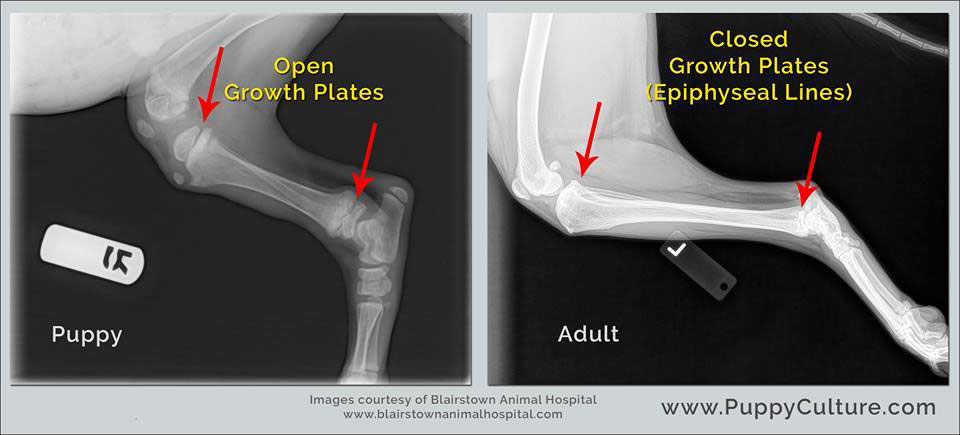
About Spay/Neuter, and how early. . .
Between Rocks and a Hard Place?
by Karen P. Christensen
Early Spay-Neuter Considerations for the Canine Athlete: One Veterinarian’s Opinion

|
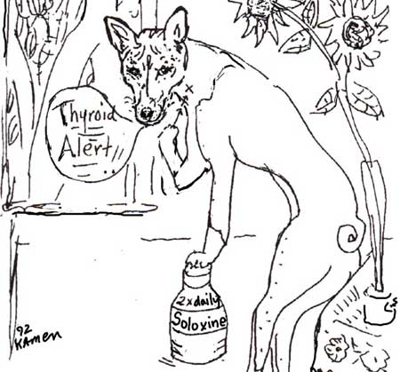
About Thyroid Testing Poem from May of 2000. "Little Timmy Sure looks skinny," Susan said one day. "He looks morose, His coat is gross-- He seems so far away." So Little Timmy Called his vet And told him of his woes The vet said, "Timmy, I can help, but you can't bite my nose." "I'll have to take Some blood from you, a whole bunch, from your neck-- Is this OK?" Tim gave a nod. "I'll send this to Jean Dodds." Jean wrote back Her lab report-- She's very quick, you know She wrote in her red pen That Timmy's thyroid was too low! So Timmy now takes little pills, He takes them twice a day, His eyes are bright, his coat's just right And now he likes to play. And so, dear friends, You listen up-- If your dog is off the mark-- Let Jean Dodds run the test for you And get your dog his spark.
the end Lisa Osenni, Hairless & Barkless in Woodstock NY{Timmy had Low Free T4s, and normal circulating antibodies. He took his Soloxine
|
|
THYROID DISEASE AND AUTOIMMUNE THYROIDITIS Contact Dr. Dodds at HEMOPET / HEMOLIFE . Introduction Hypothyroidism is the most common endocrine disorder of canines, and up to 80% of cases result from autoimmune (lymphocytic) thyroiditis. The heritable nature of this disorder poses significant genetic implications for breeding stock. Thus, accurate diagnosis of the early compensatory stages of canine autoimmune thyroiditis leading up to hypothyroidism affords important genetic and clinical options for prompt intervention and case management. Although thyroid dysfunction is the most frequently recognized endocrine disorder of pet animals, it is often difficult to make a definitive diagnosis. As the thyroid gland regulates metabolism of all body cellular functions, reduced thyroid function can produce a wide range of clinical manifestations. Many of these clinical signs mimic those resulting from other causes and so recognition of the condition and interpretation of thyroid function tests can be problematic (Table 1). Baseline Thyroid Profiles A complete baseline thyroid profile is measured and typically includes total T4, total T3, free T4, free T3, T3AA and T4AA, and can include cTSH and/or TgAA. The TgAA assay is especially important in screening breeding stock for heritable autoimmune thyroid disease. The normal reference ranges for thyroid analytes of healthy adult animals tend to be similar for most breeds of companion animals. Exceptions are the sighthound and giant breeds of dogs which have lower basal levels. Typical thyroid levels for healthy sighthounds, such as retired racing greyhounds, are at or just below the established laboratory reference ranges, whereas healthy giant breeds have optimal levels around the midpoint of these ranges. Similarly, because young animals are still growing and adolescents are maturing, optimal thyroid levels are expected to be in the upper half of the references ranges. For geriatric animals, basal metabolism is usually slowing down, and so optimal thyroid levels are likely to be closer to midrange or even slightly lower. Genetic Screening for Thyroid Disease Most cases of thyroiditis have elevated serum TgAA levels, whereas only about 20-40% of cases have elevated circulating T3 and/or T4 AA. Thus, the presence of elevated T3 and/or T4 AA confirms a diagnosis of autoimmune thyroiditis but underestimates its prevalence, as negative (non-elevated) autoantibody levels do not rule out thyroiditis. Measuring TgAA levels also permits early recognition of the disorder, and facilitates genetic counselling. Affected dogs should not be used for breeding (Table 2). 2 The commercial TgAA test can give false negative results if the dog has received thyroid supplement within the previous 90 days, thereby allowing unscrupulous owners to test dogs while on treatment to assert there normalcy, or to obtain certification with health registries such as the OFA Thyroid Registry. False negative TgAA results also can occur in about 8% of dogs verified to have high T3AA and/or T4AA. Furthermore, false positive TgAA results may be obtained if the dog has been vaccinated within the previous 30-45 days, or in some cases of nonthyroidal illness. Vaccination of pet and research dogs with polyvalent vaccines containing rabies virus or rabies vaccine alone was recently shown to induce production of antithyroglobulin autoantibodies, a provocative and important finding with implications for the subsequent development of hypothyroidism. A population study of 287,948 dogs was recently published by the MSU Animal Health Diagnostic Laboratory. Circulating thyroid hormone autoantibodies (T3AA and/or T4AA)) were found in 18,135 of these dogs (6.3%). The 10 breeds with the highest prevalence of thyroid AA from their study were: Pointer, English Setter, English Pointer, Skye Terrier, German Wirehaired Pointer, Old English Sheepdog, Boxer, Maltese, Kuvasz, and Petit Basset Griffon Vendeen. Prevalence was associated with body weight and was highest in dogs 2-4 years old. Females were significantly more likely to have thyroid AA than males. A bitch with circulating thyroid AA has the potential to pass these along to the puppies transplacentally as well as via the colostrum. Furthermore, any dog having thyroid AA may eventually develop clinical symptoms of thyroid disease and/or be susceptible to other autoimmune diseases. Thyroid screening is thus very important for selecting potential breeding stock as well as for clinical diagnosis. Thyroid testing for genetic screening purposes is less likely to be meaningful before puberty. Screening is initiated, therefore, once healthy dogs and bitches have reached sexual maturity (between 10-14 months in males and during the first anestrous period for females following their maiden heat). As the female sexual cycle is quiescent during anestrus, any influence of sex hormones on baseline thyroid function will be minimized. This period generally begins 12 weeks from the onset of the previous heat and lasts one month or longer. The interpretation of results from baseline thyroid profiles in intact females will be more reliable when they are tested in anestrus. In fact, genetic screening of intact females for other disorders such as von Willebrand disease (vWD), hip dysplasia, and wellness or reproductive checkups (vaginal cultures, hormone testing) is best scheduled during anestrus. Once the initial thyroid profile is obtained, dogs and bitches should be rechecked on an annual basis to assess their thyroid function and overall health. Generation of annual test results provides comparisons that permit early recognition of developing thyroid dysfunction. This allows for early treatment, where indicated, to avoid the appearance or advancement of clinical signs associated with hypothyroidism. Canine autoimuune thyroid disease is very similar to Hashimoto’s thyroiditis of humans, which has been shown to be associated with human major histocompatibility complex (MHC) tissue types. A similar association with canine MHC genes in hypothyroid dogs has recently been reported in Doberman Pinschers, English Setters and Rhodesian Ridgebacks, who share a rare dog leukocyte antigen (DLA) class II haplotype which contains a unique DLA-DQA1*00101 genetic determinant. While the presence of this determinant doubles the risk of a dog developing hypothyroidism, it was not found in boxers affected with thyroiditis, nor was it found in the Shih Tzu, Yorkshire Terrier, or Siberian Husky, although more studies are needed in these and other 3 susceptible breeds to establish their true status with respect to this marker DLA antigen. This exciting finding of a common genetic determinant associated with thyroid disease in several breeds hopefully will lead to development of a genetic marker test to identify affected breeding stock and allow for selective breeding to reduce disease incidence in pure-bred dogs. Polyglandular Autoimmunity Individuals genetically susceptible to autoimmune thyroid disease may also become more susceptible to immune-mediated diseases affecting other target tissues and organs, especially the bone marrow, liver, adrenal gland, pancreas, skin, kidney, joints, bowel, and central nervous system. The resulting “polyglandular autoimmune syndrome” of humans is becoming more commonly recognized in the dog, and probably occurs in other species as well. The syndrome tends to run in families and is believed to have an inherited basis. Multiple endocrine glands and nonendocrine systems become involved in a systemic immune-mediated process. This multiple endocrinopathy often occurs in patients with underlying autoimmune thyroid disease (hypo- or hyperthyroidism) and concurrent Addison’s disease, diabetes, reproductive gonadal failure, skin disease and alopecia, and malabsorption syndrome. The most common nonendocrinologic autoimmune disorders associated with this syndrome are autoimmune hemolytic anemia (AIHA), idiopathic thrombocytopenic purpura (ITP), chronic active hepatitis, and immune-complex glomerulonephritis (systemic lupus erythematosus; SLE). The most commonly recognized polyglandular endocrinopathy of dogs is Schmidt’s syndrome (thyroiditis and Addison’s disease). Examples of breeds genetically predisposed to this disorder include the Standard Poodle, Old English Sheepdog, Bearded Collie, Portuguese Water Dog, Nova Scotia Duck Tolling Retriever, and Leonberger, although any breed or mixed breed can be affected. Our study cohort of 162 cases of autoimmune blood and endocrine disorders in Old English Sheepdogs (1980-1989) included 115 AIHA and/or ITP, 99 thyroid disease, 23 Addison’s disease, 7 vaccine reactions, 3 SLE, 2 diabetes, 1 rheumatoid arthritis and 1 hypoparathyroidism. The group comprised 110 females (15 spayed) and 52 males (3 neutered). Seven of the most recent 103 cases had two or more endocrine disorders, and 101 of the 108 cases where pedigrees were available showed a familial relationship going back several generations. Data from surveying the Bearded Collie breed reported 55 hypothyroid, 17 Addison’s disease, and 31 polyglandular autoimmunity (5 were hypothyroid). The Hemopet/Hemolife Test Request Form and Instructions can now be filled out online. |
